41 pearson heart diagram
Heart Diagram Answer Key.indd Author: uweb Created Date: 5/20/2009 11:07:16 PM ... 86 Chapter 4 Anatomy and Physiology of the Skeletal System bone marrow bones joints ligaments (LIG-ah-ments) skeleton Each bone in the human body is a unique organ that carries its own blood
The Heart - Science Quiz: Day after day, your heart beats about 100,000 times, pumping 2,000 gallons of blood through 60,000 miles of blood vessels. If one of your organs is working that hard, it makes sense to learn about how it functions! This science quiz game will help you identify the parts of the human heart with ease. Blood comes in through veins and exists via arteries—to control the ...

Pearson heart diagram
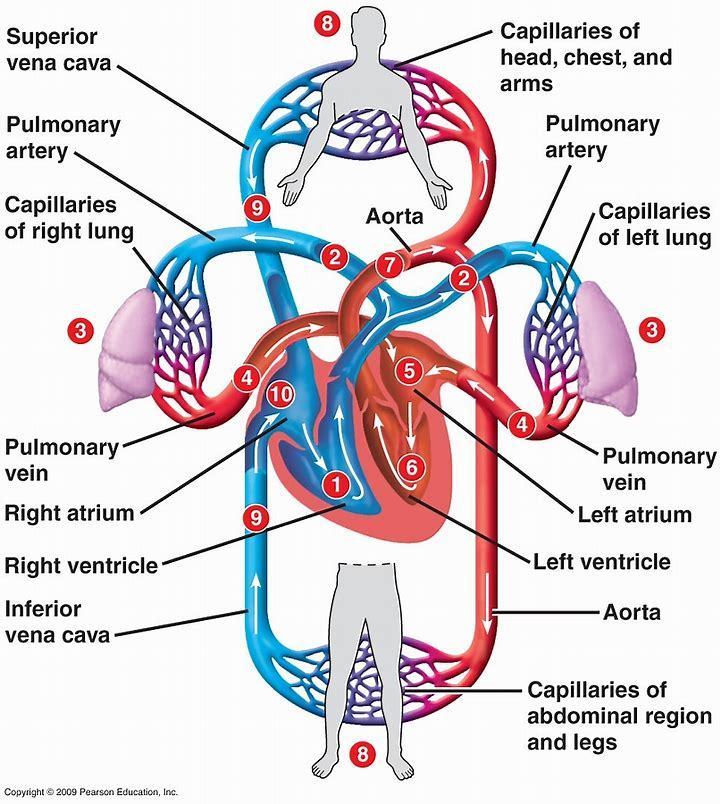
The modest size and weight of the heart belie its incredible strength and endurance. About the size of a fist, the hollow, cone-shaped heart has a mass of 250 to 350 grams—less than a pound (Figure 18.2). Figure 18.1 The systemic and pulmonary circuits. The right side of the heart pumps blood through the pulmonary circuit and There are 4 chambers, labeled 1-4 on the diagram below. To help simplify things, we can convert the heart into a square. We will then divide that square into 4 different boxes which will represent the 4 chambers of the heart. The boxes are numbered to correlate with the labeled chambers on the cartoon diagram. Live worksheets > English. Label parts of the heart. Drag and drop the labels to the correct parts indicated on the heart diagram. ID: 832107. Language: English. School subject: Biology. Grade/level: GCSE. Age: 12-18. Main content: Label parts of the heart.
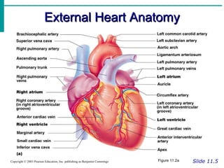
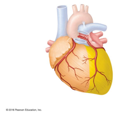
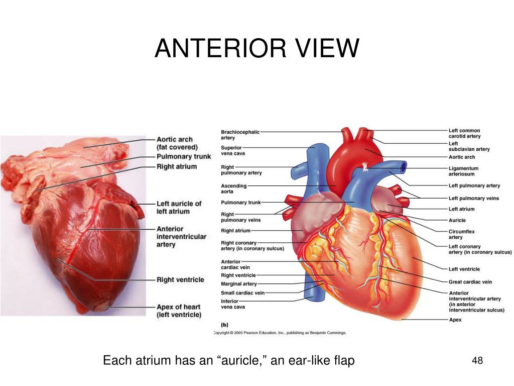
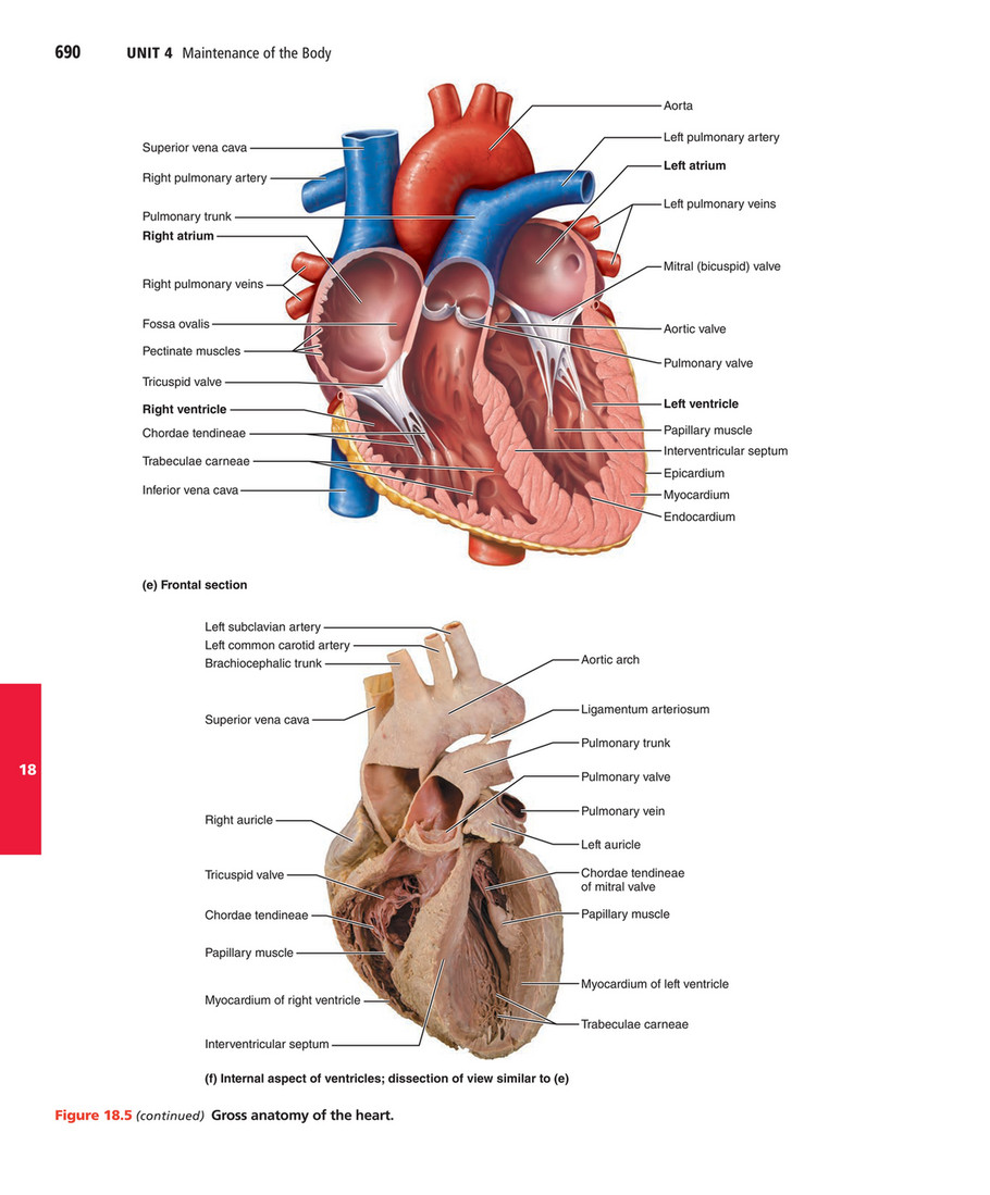
Pearson heart diagram. Mastering A&P. Assignable content and assessments help your students quickly master concepts, keeping them engaged and on track. Foster student engagement and collaborative learning. Adaptive follow-up assignments for Mastering™ offer a truly personalized learning experience with targeted homework help that adapts in real time. (a) Anterior view of the external heart C' 2019 Pearson Education. Aort'c arch Ligamentum arteriosum Left pulmonary artery Left pulmonary ve ns Auricle of left atrium Circumflex artery Left coronary artery (in atrioventricular sulcus) Great cardiac vein Left ventricle Anterior interventricular artery (in anterior interventricular sulcus) Apex The anatomy of the eye is fascinating and this quiz game will help you memorize the 12 parts of the eye with ease. 84 images of Heart Diagram Unlabeled. Module 1 Labeled Diagram Of The Eye Diagram Of The Eye Dot Worksheets Diagram. Label Parts Of The Human Eye. Eye Anatomy 1 Illustration Photo In 2021 Eye Anatomy Eye Anatomy Diagram Human Eye ... ©2016 Pearson Education Ltd. 1/1/1/1/1/1/ *P45629A0132* Biology Unit: 4BI0 Science (Double Award) 4SC0 ... The diagram shows what the student observes through the microscope. One cell has ... 7 The diagram shows the structure of the human heart. Structure C Side X Side Y
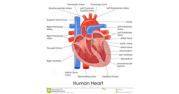
Pearson Edexcel International GCSE. 2 *P44262A0216* Answer ALL questions. 1 (a) The diagram shows the human circulatory system. (i) Draw an arrow in each of the blood vessels A, B, C and D to show the direction of ... and heart rate. The graphs show the results of the investigation. arterial blood pressure in mm of mercury It consists of the heart, which is a muscular pumping device, and a closed system of vessels called arteries, veins, and capillaries. As the name im-plies, blood contained in the circulatory system is pumped by the heart around a closed circle or circuit of vessels as it passes again and again through the various circulations of the body (on p ... ©2012 Pearson Education Ltd. 1/1/1/1/ Turn over Edexcel International GCSE. 2 *P40128A0228* ... The diagram shows a section through the human heart. The correct direction for the flow of blood in the heart is from chamber (1) A to B B to C ... The diagram shows the genotypes of a father and a mother. Diagram of Heart. The human heart is the most crucial organ of the human body. It pumps blood from the heart to different parts of the body and back to the heart. The most common heart attack symptoms or warning signs are chest pain, breathlessness, nausea, sweating etc. The diagram of heart is beneficial for Class 10 and 12 and is frequently ...
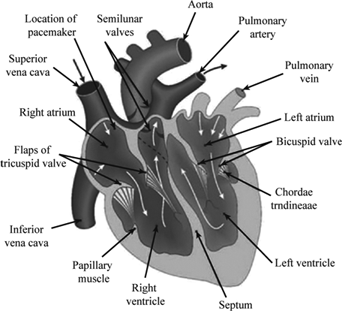
The heart weighs less than 0.5 kg (1 lb) in a nor - mal healthy adult. Two-thirds of the heart mass lies to the left of the sternum; the upper base lies beneath the second rib, and the pointed apex is approximate with the fifth intercostal space, midpoint to the clavicle (Figure 29-1 •). The heart is covered by the pericardium, a double ... The diagram shows a section through a human heart as positioned in the body. 2 Pearson Leve 3 ationas in eath and ocia are - nit 3 - Fina ape Assessent aterias - ssue 1 - oveber 21 earson ducation Liited 21 Selecting or hovering over a box will highlight each area in the diagram. In this interactive, you can label parts of the human heart. Drag and drop the text labels onto the boxes next to the heart diagram. If you want to redo an answer, click on the box and the answer will go back to the top so you can move it to another box. 1. the heart is an essential pumping organ in the cardiovascular system where the right heart pumps deoxygenated blood (returned from body tissues) to the lungs for gas exchange, while the left heart pumps oxygenated blood (returned from the lungs) to tissues cells for sustaining cellular respiration. 2. Attached to the heart is blood vessels ...
Pearson Edexcel International GCSE. 2 *P45936A0228* DO NOT WRITE IN THIS AREA DO NOT WRITE IN THIS AREA DO NOT WRITE IN THIS AREA Answer ALL questions. 1 For each of the questions (a) to (j), choose an answer, AB, C or D and put a cross in the ... The diagram shows a heart without any valves being drawn. (i) Name the blood vessels A, B, C and D
Basilic vein . Brachial vein . Median cubital vein . Dorsal venous arch . Small saphenous vein . Digital veins . Great saphenous vein . Ulnar vein
©2016 Pearson Education Ltd. 1/1/3/1/ *P46931A0124* Biology Advanced Subsidiary ... The diagram below shows the structure of a molecule of glucose. ... When the human heart contracts, blood from the left ventricle enters the aorta.
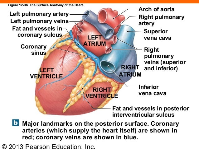
Section 1: Structure of the Heart Learning Outcomes (continued) 18.5 Describe the major vessels supplying the heart, and cite their locations. 18.6 Trace blood flow through the heart, identifying the major blood vessels, chambers, and heart valves. 18.7 Describe the relationship between the AV and semilunar valves during a heartbeat.
heart. These lightly striated muscles function more efficiently through the use of intercalated discs. This involuntary muscle type is found in the walls of hollow organs and vessels as well as respiratory airways. These nonstriated muscles contract and relax and help perform peristalsis and maintain the diameter of blood vessels and airways.
Title: Venn Diagram Author: Connections Education Subject: Discussion Guidelines and Rubric Keywords "connections, education, graphic, organizer, venn, diagram"
* Zoe recorded the heart rates, in beats per minute, of each of 15 people. Zoe then asked the 15 people to walk up some stairs. She recorded their heart rates again. She showed her results in a back-to-back stem and leaf diagram. Compare the heart rates of the people before they walked up the stairs
Describe the location of the heart in the body, CPF|KFGPVKH[ KVU OCLQT CPCVQOKECN CTGCU QP CP appropriate model or diagram. Size, Location, and Orientation The modest size and weight of the heart give few hints of its incredible strength. Approximately the size of a person's fist, the hollow, cone-shaped heart weighs less than a pound.
Live worksheets > English. Label parts of the heart. Drag and drop the labels to the correct parts indicated on the heart diagram. ID: 832107. Language: English. School subject: Biology. Grade/level: GCSE. Age: 12-18. Main content: Label parts of the heart.
There are 4 chambers, labeled 1-4 on the diagram below. To help simplify things, we can convert the heart into a square. We will then divide that square into 4 different boxes which will represent the 4 chambers of the heart. The boxes are numbered to correlate with the labeled chambers on the cartoon diagram.
The modest size and weight of the heart belie its incredible strength and endurance. About the size of a fist, the hollow, cone-shaped heart has a mass of 250 to 350 grams—less than a pound (Figure 18.2). Figure 18.1 The systemic and pulmonary circuits. The right side of the heart pumps blood through the pulmonary circuit and









Comments
Post a Comment